Labs & Equipment

Cell Biology Lab

All experiments related to cell culture are carried out in this laboratory. It has a laminar flow cabinet, an incubator, two microscopes of phase contrast, a thermal bath, and different types of centrifuges.

Molecular Biology Lab

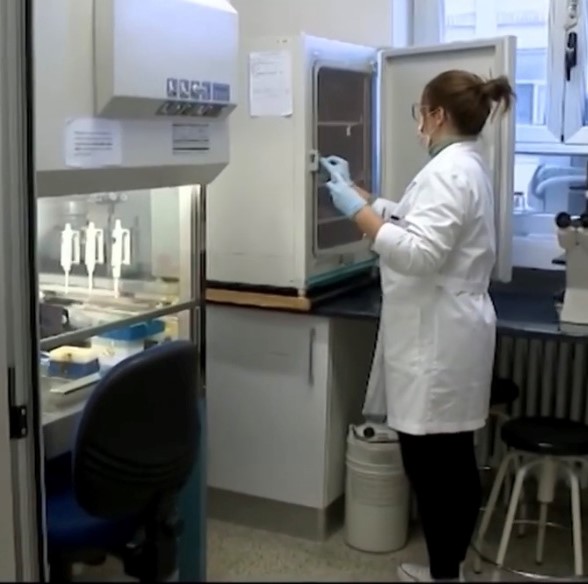
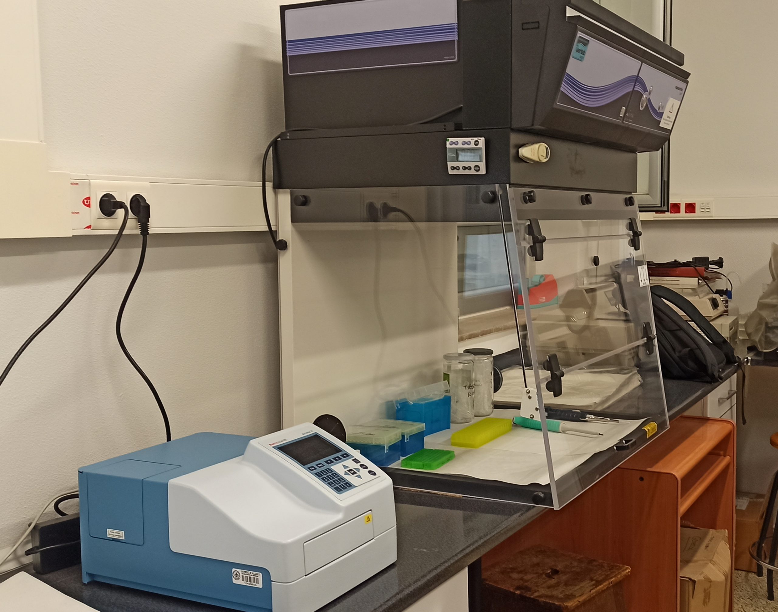
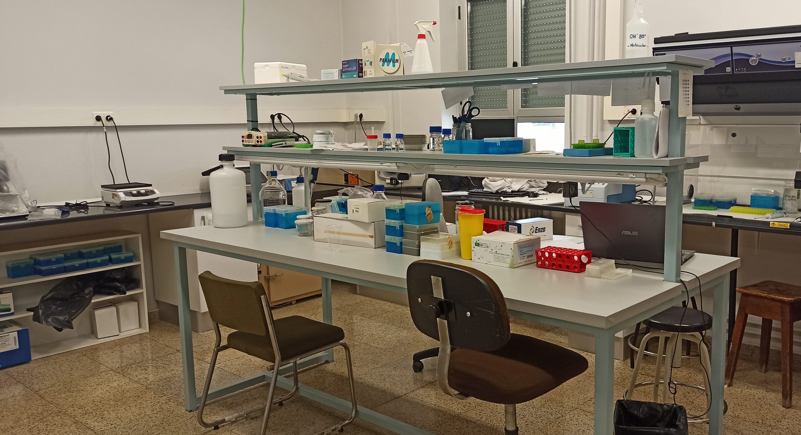
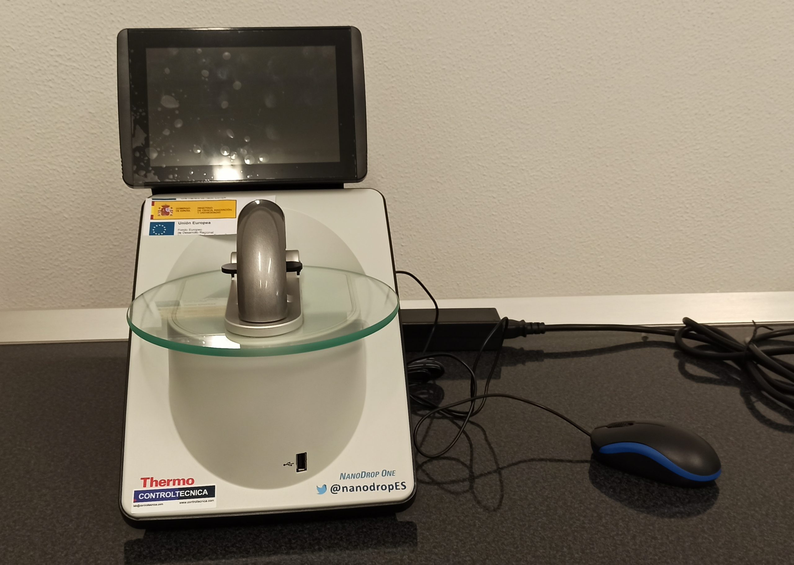
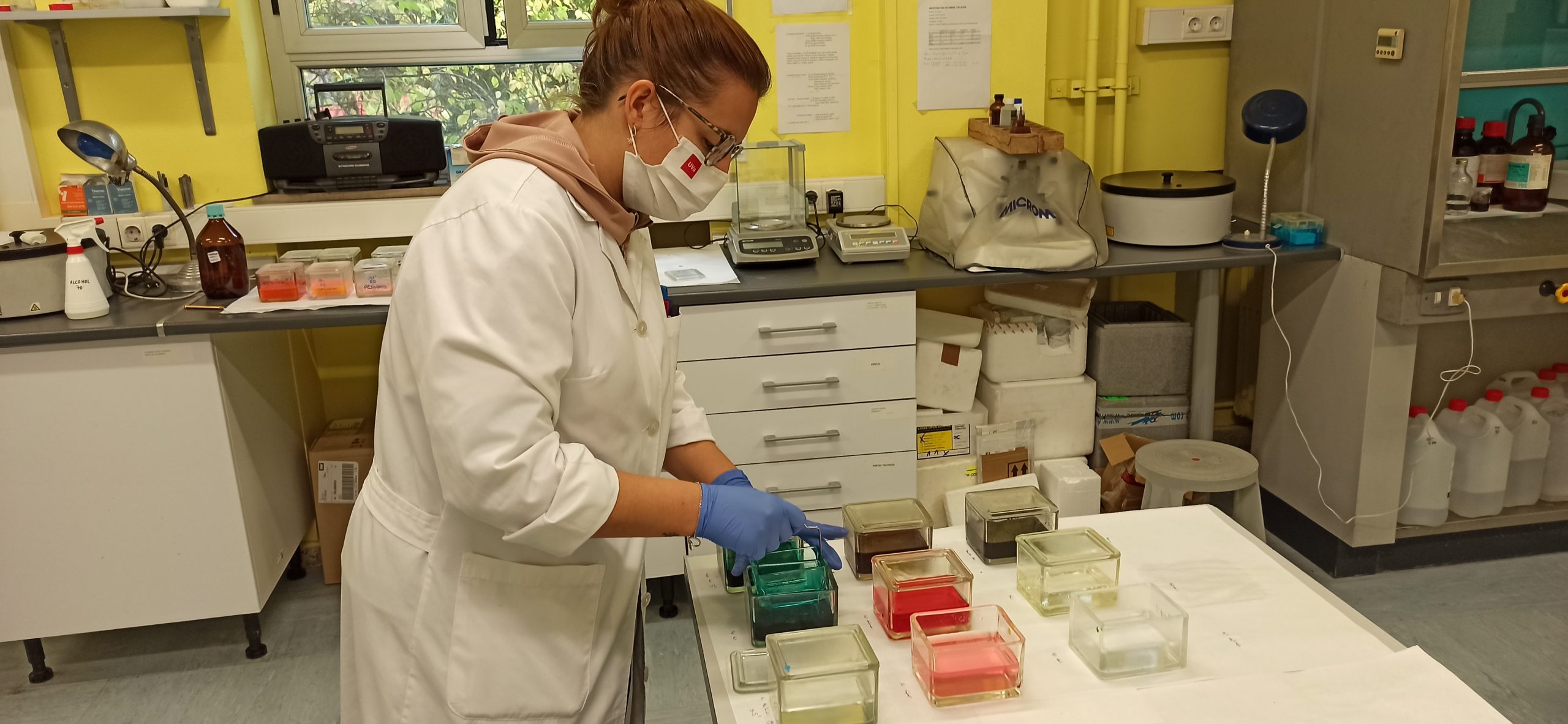
In the Molecular Biology laboratory, we carry out all the sample processing for subsequent molecular analysis. This laboratory has a cabinet where RNA and protein extraction is carried out, a thermal cycler, a nano drop, a plate reader for ELISA determinations and all the material necessary to assess gene expression by RT-PCR and protein expression by Western-blot.

Histology Lab

In the Histology lab we carry out all histological processing of biological samples. For this purpose, this laboratory has all the necessary equipment including an automatic processor, a paraffin station, cutting apparatus (microtome and cryostat) and different batteries to perform different staining and histochemical techniques.

Spectroscopy Lab

This laboratory is equipped with the necessary instruments to measure the transmittance of ophthalmic or intraocular lenses and “ex vivo” corneas. It is also able to measure the spectral distribution of irradiance of commonly used lamps, LEDs, or specific illuminants. It also has a SAFAS spectrophotometer, used to obtain measurements of the neutral density filters used in the experiments.

Radiometry, Vision, and Illumination Lab

This laboratory has two devices with different but complementary research lines:

  • A Maxwellian double vision optical device capable of carrying out this type of psychophysical measurements, with precision of 0.1 logarithmic units in the threshold contrast measurement and temporal resolution of the order of a millisecond. It consists of a laser, lenses, neutral density filters, diaphragms, shutters, beam splitters and pupillometer. This system allows the measurement of threshold contrast, visual reaction time, glare recovery time, as well as the control and determination of pupil diameter. All this under different conditions of illumination, spectral composition, sizes, and presentation times of both the stimulus and the adaptation field. The software that controls the computer for performing the tests, as well as the luminance meter for measuring luminance, is also located on the room.
  • A two-channel photostimulator consists of ultraluminescent LEDs with interferential filters, fibre-optic harnesses, spatial homogenisers, photographic objectives, a prism, a photometric cube, lenses, and diaphragms. The quasi-monochromatic (10 nm) spectral emission of each LED is close to the peak of maximum spectral absorption of each of the photoreceptors in the retina of a standard observer. In this case the shuttering of the beams and the intensity control is electronic, and computer controlled. The device allows not only the measurement of threshold contrast, reaction time and estimation of the underlying adaptation mechanisms, but also the application of the ‘silent substitution’ technique for the isolated study of photoreceptors and post-receptoral pathways.